Definition and Branches of Anatomy
MBBS study notes with upper limb applications
Anatomy describes the structures of the body and the relationships that make those structures clinically meaningful. These notes organize the subject by scale and method, then connect upper-limb anatomy with evolution, examination, and common injury patterns.
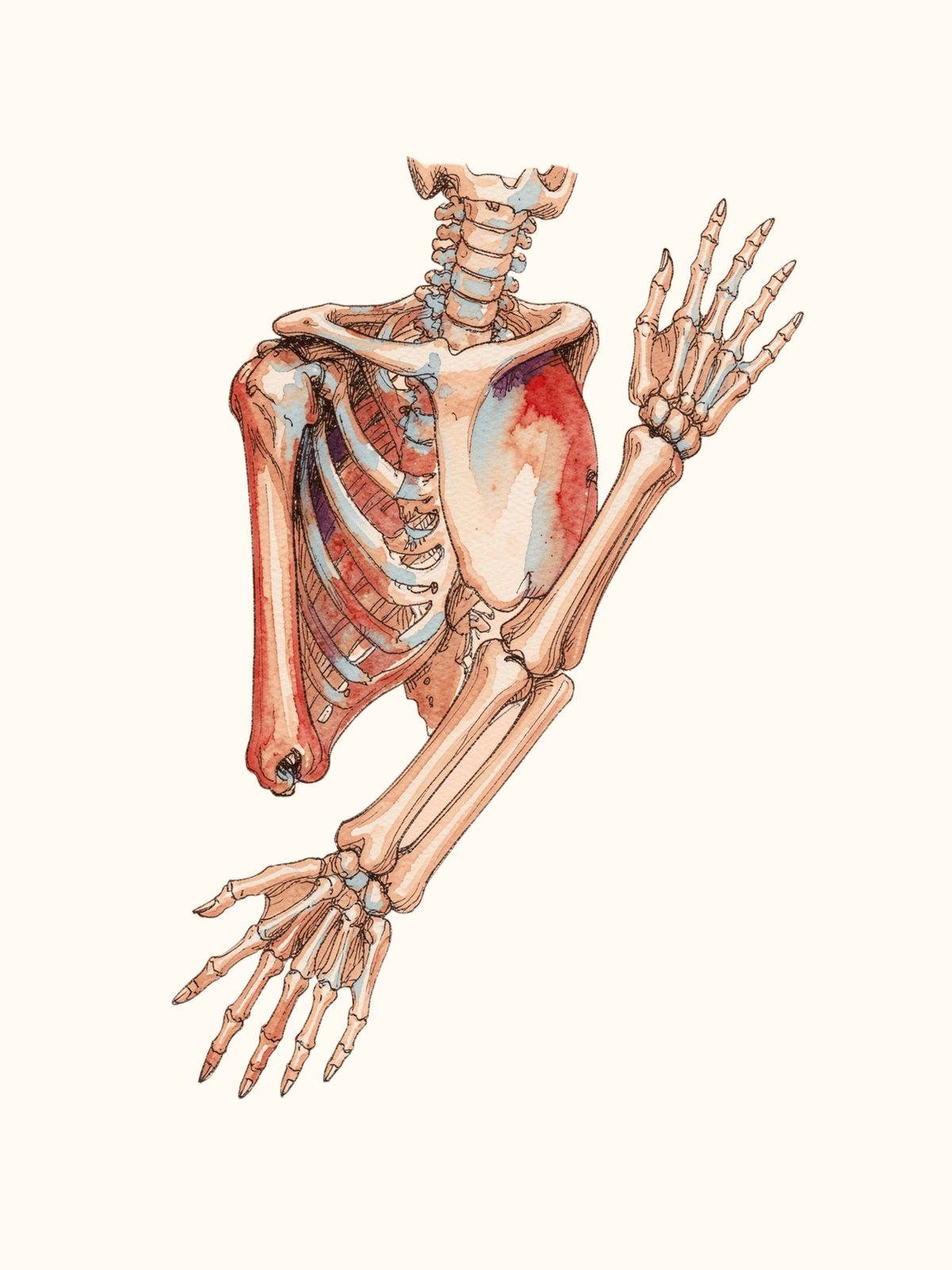
Upper limb from the shoulder girdle to the hand. Illustrative AI-generated image; use a standard atlas for exact anatomical detail.
Definition
Anatomy (Greek: ana = up; tome = cutting) is the scientific study of the structure of the human body and the relationships among its parts.
- Scope. Anatomy describes form, location, relations, blood supply, nerve supply, lymphatics, development, and function of body structures.
- Goal. It provides the structural basis for understanding physiology, pathology, clinical examination, imaging, and surgery.
Major branches of anatomy
Gross or macroscopic anatomy
|
Branch
|
Focus and use
|
|
Systemic anatomy
|
Studies the body by organ systems, such as skeletal, muscular, and cardiovascular systems. Useful for organizing theory.
|
|
Regional anatomy
|
Studies all structures in one region, from skin to deep structures, such as the axilla, abdomen, or head and neck. Useful in dissection, clinics, and surgery.
|
|
Surface anatomy
|
Uses body-surface landmarks to locate deeper structures. Supports clinical examination and procedures.
|
|
Radiological or imaging anatomy
|
Interprets structures on X-ray, ultrasound, CT, MRI, and angiography. Supports diagnosis and intervention.
|
|
Cross-sectional anatomy
|
Studies structures in transverse, coronal, and sagittal planes, improving correlation with imaging.
|
|
Living anatomy
|
Studies structures in a living person through inspection, palpation, percussion, auscultation, and functional tests.
|
|
Clinical, surgical, or applied anatomy
|
Applies anatomical facts to clinical problems such as injuries, nerve lesions, and surgical approaches.
|
|
Functional anatomy
|
Relates structure to function, including how shape or attachment produces movement.
|
|
Topographic anatomy
|
Studies spatial relationships within a region, including planes, triangles, and spaces.
|
Microscopic anatomy
|
Branch
|
Focus
|
|
Histology
|
Tissues under the light microscope and organ microstructure. Includes epithelial, connective (cartilage, bone, blood), muscle, and nervous tissues.
|
|
Cytology
|
Individual cells, including their shape, organelles, and inclusions. It forms a foundation for pathology.
|
|
Ultrastructure
|
Subcellular architecture studied by electron microscopy, such as basement membrane and mitochondrial cristae.
|
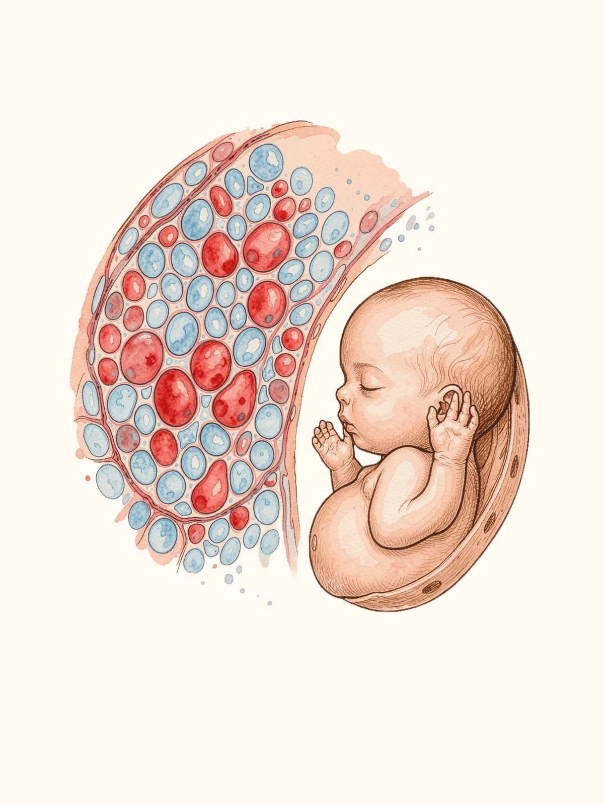
Illustrative plate linking tissue-scale anatomy with development. AI-generated schematic; not a diagnostic or histology reference.
Developmental anatomy
|
Branch
|
Focus
|
|
Embryology
|
Prenatal development from gametogenesis and fertilization through cleavage, gastrulation, organogenesis, and the fetal period.
|
|
Teratology
|
Congenital anomalies, including causes such as genetic factors and teratogens, mechanisms, and prevention.
|
|
Postnatal growth and aging
|
Changes after birth, including growth plates, secondary ossification, and involution.
|
Specialized or focused branches
|
Branch
|
Focus and relevance
|
|
Neuroanatomy
|
CNS, PNS, and ANS, including gross structures, pathways, and nuclei. Important in neurology and neurosurgery.
|
|
Comparative anatomy
|
Compares structures across species to support evolutionary and functional understanding.
|
|
Anthropometry and physical anthropology
|
Studies body measurements and variation, with forensic relevance.
|
|
Molecular anatomy
|
Studies structure at the protein and DNA levels, linking anatomy with genetics and precision medicine.
|
Quick exam comparisons
|
Comparison
|
Key distinction
|
|
Systemic vs regional
|
Systemic anatomy follows organ systems. Regional anatomy studies all structures within one area.
|
|
Surface vs living
|
Surface anatomy focuses on landmarks. Living anatomy studies the person through examination and functional tests.
|
|
Imaging vs cross-sectional
|
Imaging anatomy is organized by modality. Cross-sectional anatomy describes structures in planes and helps orient images.
|
|
Embryology and teratology
|
The timing of an insult relates to the type of anomaly. Organogenesis is the period of highest risk.
|
Evolution of the upper limbs
Forelimbs of vertebrates share a common structural plan. In the notes below, compare how that plan is modified for different functions.
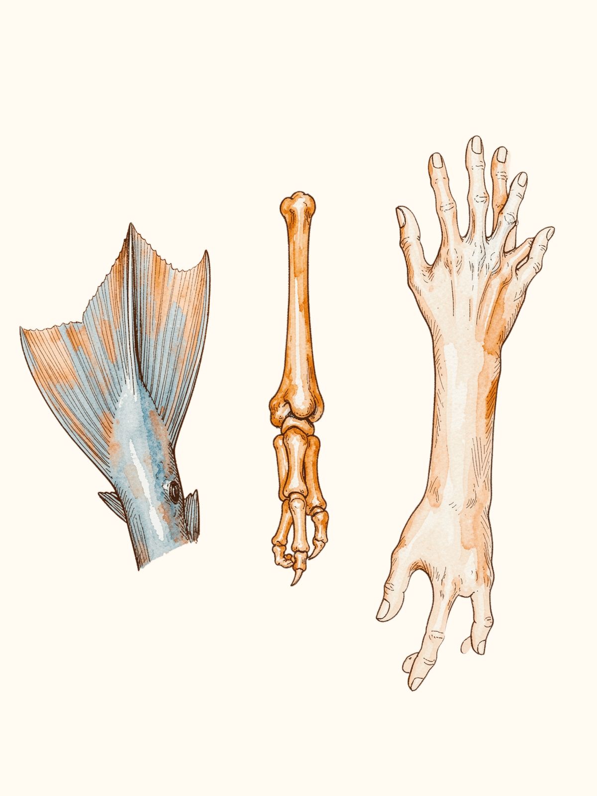
Comparative forelimb illustration. The image is schematic; rely on anatomical references for detailed bone homologies.
|
Group
|
Main adaptation and features
|
|
Fish
|
Paired pectoral and pelvic fins supported by cartilaginous or bony girdles.
|
|
Amphibians (e.g., frog)
|
Limbs adapted for land locomotion. The skeleton includes humerus, radius, ulna, carpals, metacarpals, and phalanges. The palm is directed backward.
|
|
Reptiles (e.g., lizard)
|
Limbs support creeping or crawling. The palm is directed forward.
|
|
Birds
|
Forelimbs are modified into wings for flight. Skeletal elements remain specialized, with elongated metacarpals and fused digits.
|
|
Mammals, including humans
|
Limbs support grasping, manipulation, and locomotion. Primates have a prehensile hand; an opposable thumb enables precision grip. The shoulder has the greatest range of motion. In humans, the upper limb mainly supports skill and manipulation rather than locomotion.
|
Study of upper-limb anatomy
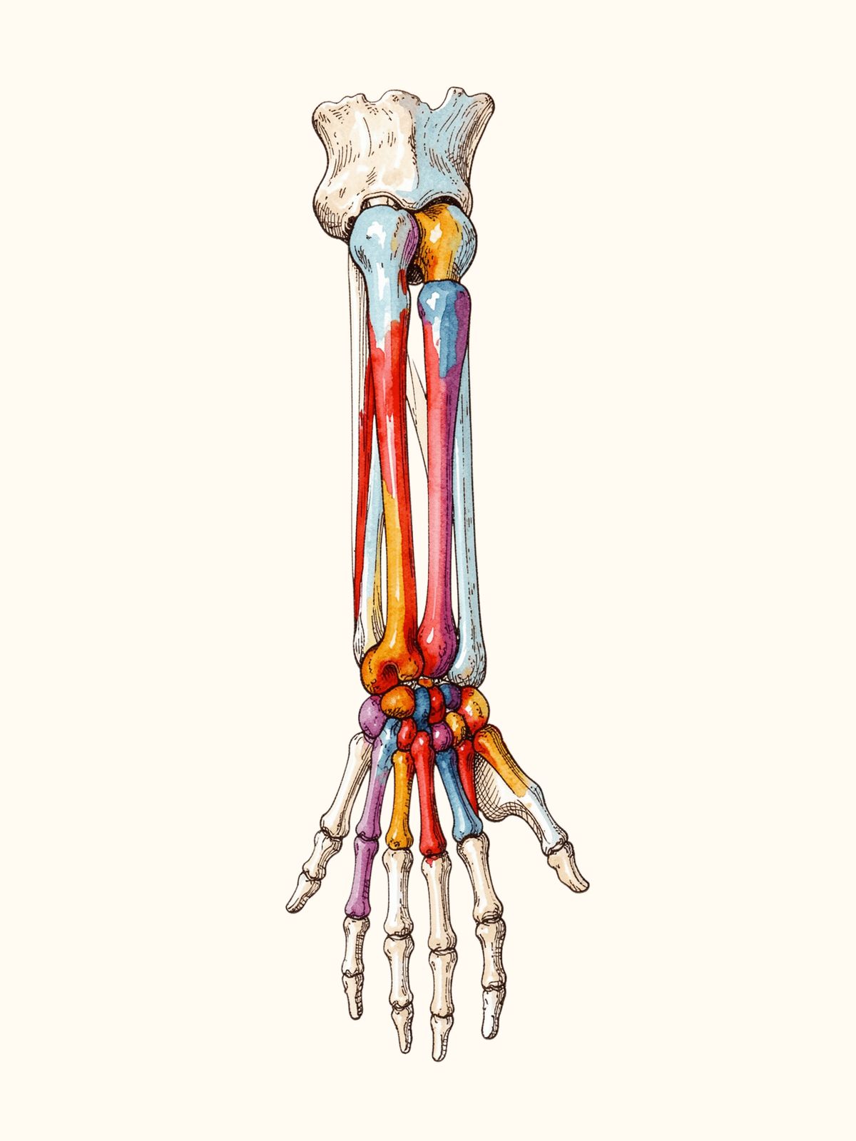
Upper-limb skeleton shown from the shoulder girdle to the fingertips. Illustrative AI-generated image.
|
Method
|
What to study
|
|
Osteology
|
Bones: shape, features, ossification centers, attachments, relations, and applied importance.
|
|
Arthrology
|
Joints and ligaments: types, movements, stability, and applied anatomy.
|
|
Myology
|
Muscles: origin, insertion, nerve supply, actions, and applied anatomy.
|
|
Angiology
|
Arteries, veins, lymphatic drainage, and clinical importance.
|
|
Neurology
|
Peripheral nerves, plexuses, sensory and motor distribution, and nerve injuries.
|
|
Surface anatomy
|
Markings of important structures on the body surface for examination and surgery.
|
|
Radiological anatomy
|
Radiographs, CT, and MRI to identify bones, joints, and organs.
|
|
Embryology
|
Development of the upper limb and the basis of congenital anomalies.
|
|
Comparative and evolutionary anatomy
|
Species comparisons that help explain structural modifications.
|
Arm and forearm compartments
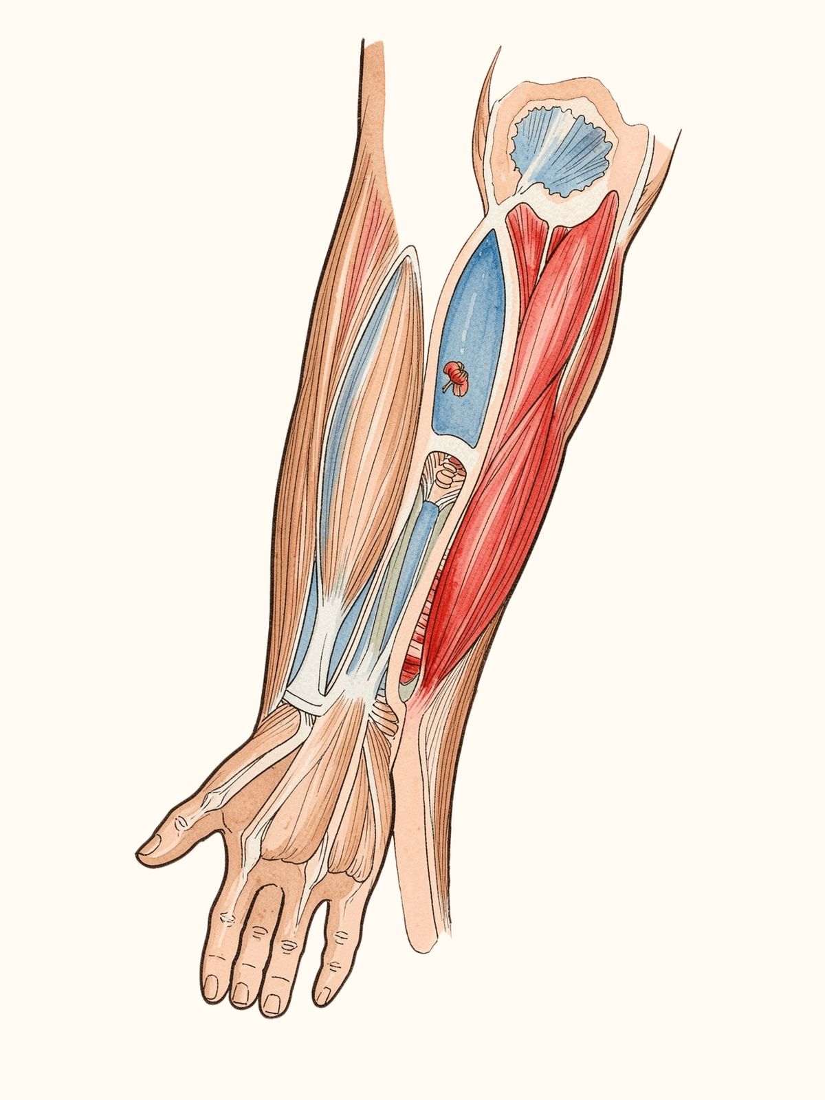
Schematic cross-sections of the upper arm and forearm compartments. Confirm detailed muscle and nerve relationships with an atlas.
|
Region and compartment
|
Key muscles or nerve
|
|
Arm, anterior
|
Biceps brachii; musculocutaneous nerve.
|
|
Arm, posterior
|
Triceps; radial nerve.
|
|
Forearm, anterior
|
Median and ulnar nerves.
|
|
Forearm, posterior
|
Radial nerve.
|
Frequently asked questions
1. Why is the human upper limb more mobile than the lower limb?
It is not primarily concerned with weight-bearing. The highly mobile shoulder joint permits a wide range of movement.
2. Which features make the human hand prehensile?
The opposable thumb, long mobile fingers, and well-developed intrinsic muscles.
3. Which arm and forearm compartments are clinically important?
Arm: anterior compartment with biceps and the musculocutaneous nerve; posterior compartment with triceps and the radial nerve. Forearm: anterior compartment with median and ulnar nerves; posterior compartment with the radial nerve.
4. Which methods are used to study upper-limb anatomy?
Dissection, osteology, radiology, surface anatomy, and embryological correlation.
5. What common upper-limb injury sites are important for exams?
Clavicle fracture; surgical neck of humerus with axillary nerve injury; midshaft humerus with radial nerve injury; supracondylar humerus with brachial artery injury; and scaphoid fracture with risk of avascular necrosis.
Common injury correlations
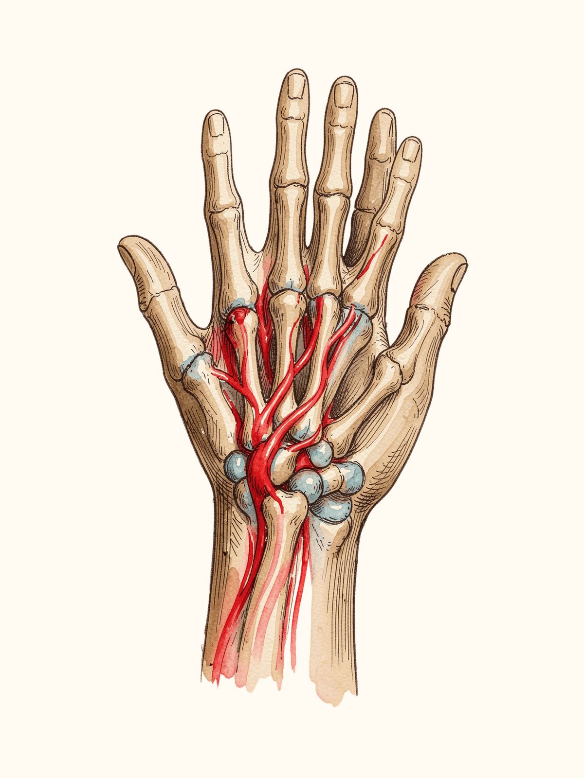
Scaphoid and its blood-supply direction shown schematically. Use an atlas or radiology reference for precise anatomy.
|
Injury site
|
Important association
|
|
Clavicle
|
Fracture
|
|
Surgical neck of humerus
|
Axillary nerve injury
|
|
Midshaft humerus
|
Radial nerve injury
|
|
Supracondylar humerus
|
Brachial artery injury
|
|
Scaphoid
|
Avascular necrosis risk
|
 Continue with Google